CLOSE X

Blog

Postnatal evaluation and management of a case of suspected skeletal dysplasia

Introduction: Skeletal dysplasia are a heterogenous group of disorders which affect the natural development of skeletal system and manifest as abnormalities of limbs, chest, skull or spine. These are genetic diseases which result in abnormal formation of chondro-osseous tissues. Their prevalence varies from 1:5000 to 1:3000 births with such wide variation primarily because of incomplete evaluation of still births and early neonatal deaths in several studies (1). These disorders contribute to a significant proportion of still births and early neonatal deaths, however several of them have better prognosis with survival well into adulthood. They have a varied clinical presentation and can be diagnosed at various ages due to this variable presentation (2). A comprehensive knowledge of these disorders is required for timely suspicion, complete examination, appropriate testing and diagnosis. Early and correct diagnosis is essential as most of these cases are lethal and a diagnosis helps in subsequent pregnancies.

Clinical presentation: Skeletal dysplasias may present in either of the following four presentations: (I) Still birth with antenatal suspicion (growth retardation, short bones, vertebral and facial anomalies) with/without antenatal diagnosis (II) still birth undiagnosed with postnatal findings of shorening, limb, chest or facial anomalies (III) neonate with shortening of trunk and/or limbs, facial anomalies, respiratory insufficiency, fractures (IV) infancy to adulthood with short stature and limb or spine deformities, fractures, eye/ear/dental abnormalities.

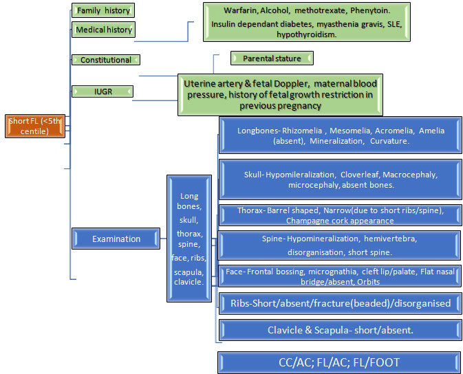
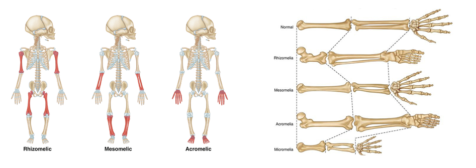
Clinical evaluation of a case of suspected skeletal dysplasia: Skeletel dysplasia is a very broad group involving host of disorders. The ones which present during neonatal period are listed in table 1 along with their features. A complete head to toe examination is required to find out all the abnormalities which can help in diagnosis. Skull should be examined for shape (barchycephaly, clover leaf, craniosynostosis), facial abnormal features like hypo/hypertelorism, cleft lip/palate, dentition anomalies, thorax should be examined for size, signs of respiratory insufficiency, spine should examined for kyphoscoliosis, limbs examined for shortening and contractures, site of shortening like proximal (rhizomelia), middle (mesomelia), distal (acromelia). Femur length-foot length ration less than 1 suggests skeletal dysplasia. Table 2 illustrates these clinical features along with their associated causes.

Radiological evaluation of a case of suspected skeletal dysplasia: A comprehensive and detailed skeletal survey is essential in aiding in diagnoses (3,4). Survey must include AP & lateral views of skull and thoracolumbar spine, AP views of chest, pelvis, one upper and lower limb, left hand (for bone age). The axial and appendicular skeleton must be properly examined for abnormalities like workman bones, thick skull, degree of ossification, platyspondyly (flat vertebral body), coronal clefts (radiolucent band running through at least one vertebral body), abnormal vertebral bodies like central beaking in Morquio’s disease and posterior hump shaped in SEDT. In appendicular skeleton, the site of affection (epiphyses, metaphases or diaphyses), type of limb shortening (rhizomelia, mesomelia, acromelia). The aetiologies associated with these findings are elaborated in table 2.

Postnatal management: After the delivery of a case of suspected skeletal dysplasia, initial stabilisation is very crucial which is to be followed by multi-disciplinary approach to diagnosis and ongoing care. Table 3 depicts the summarised care of such a baby. It is very essential to obtain samples for genetic tests for diagnosis as many of these disorders have poor prognosis and antenatal diagnosis in future pregnancies is crucial.

Conclusion: Skeletal dysplasia is a group of disorders with varied presentation and a strong clinical suspicion is necessary for timely diagnosis as outcomes are not very good. A multidisciplinary team approach is necessary to ensure a proper quality of life to survivors. Appropriate timely antenatal diagnosis at this point is primarily the mode of management. Future research on gene therapy and other treatments are required for ensuring better survival and quality of life to these individuals.

Category Disease name Clinical and radiologic features Genes involved, inheritance and prognosis
FGFR3 group Thanatropic dysplasia
  • Narrow thorax with pulmonary hypoplasia
  • Short extremeties, normal trunk length
  • Cloverleaf skull, telephone receiver femurs
  • 4p16
  • AD
  • Lethal
Achondroplasia
  • Short extremeties, rhizomelia, normal trunk lengt
  • Macrocephaly, hydrocephalus
  • Trident hand, Hypotonia
  • Foramen magnum stenosis
  • Recurrent otitis media
  • Normal intelligence
  • 4p16
  • AD
  • Excellent
Hypochondroplasia Similar to achondroplasia, milder severity, later onset in second decade
  • 4p16, AD
  • Excellent
Type II collagen group Achondrogenesis type II
  • Hydrops, short trunk, prominent abdomen
  • Severe limb shortening
  • Severely retarded bone ossification
  • 12q 13 (COL 2A1)
  • AD, Lethal
Hypochondrogenesis
  • Similar to achondrogenesis but milder shortening and better ossification
  • Most die within first 3 months from respiratory insufficiency
  • 12q 13 (COL 2A1)
  • AD, poor
Kniest dysplasia
  • Thoracic kyphoscoliosis, lumbar lordosis
  • Platyspondyly, anterior vertebral wedging, coronal clefts
  • Flat mid face, depressed nasal bridge, cleft palate
  • Joint contractures, delayed ossification and deformation of epiphyses
  • Myopia, retinal detachement
  • Chronic otitis media, hearing loss
  • 12q 13 (COL 2A1)
  • AD
  • Fair to good
Spondyloepiphyseal dysplasia congenita
  • Similar to Kniest dysplasia but milder
  • Normal long bones
  • Coronal clefts absent
  • 12q 13 (COL 2A1), AD
  • Fair to good
Stickler syndrome
  • Midface hypoplasia, small upturned nose, cleft palate, Pierre Robin sequence
  • Joint hyper mobility
  • Vertebral coronal clefts, widened epiphyses
  • Myopia, cataracts, hearing loss
  • 12q 13 (COL 2A1),
  • AD
  • Good
Short rib dysplasia group Ellis van Crevald syndrome
  • Narrow thorax, dental anomalies
  • Polydactyly, rhizomelia, hypoplastic nails
  • Congenital heart disease
  • 4p 16 (EVC1&2), AR
  • Fair to good
Asphyxiating thoracic dysplasia (Jeune)
  • Narrow thorax with respiratory insufficiency
  • Polydactyly, short hands and feet
  • Metaphysical irregularities, short middle & distal phalanges
  • Chronic kidney disease, pancreatic and hepatic fibrosis
  • Hirschsprung disease, multiple gingival frenulae, hydrocephalus
  • 3q 24-26 (IFT80), 15q 13
  • AR
  • Fair
Decreased bone density group Osteogenesis imperfecta type I-IX
  • Type II and III evident at birth, others variable age group presentation frm infancy to adulthood
  • Type II most severe with multiple fractures and high lethality, others with variable fracture rates and short stature severity
  • Blue sclera in types I to III, hearing loss in types I and III
  • I, IV,V: AD
  • II & III: AD or AR
  • VI-IX: AR
  • I to IV: COL1A1 or 2
Defective mineralization group Hypophosphatasia

  • Perinatal lethal
  • Infantile
  • Severe shortening, respiratory failure, seizures
  • Undermineralization, rachicitic metaphases, craniosynostosis
  • Short stature, premature loss of deciduous teeth, symptomatic hypercalcemia
  • 1p36 (TNAP), AR, lethal
  • 1p36 (TNAP), AR, variable

Table 1: Skeletal dysplasia which present during neonatal period. Abbreviations: AD autosomal dominant, AR autosomal recessive.

Part of body Clinical or radiological feature Possible ethology
SKULL Macrocephaly/ Microcephaly TD, achondroplasia, JLS/ CDP
Irregular skull shape TD (cloverleaf), CDP, JLS
Hydrocephalus CD, Oi type 2, osteoporosis
Normal size

  • Partial absent bones
  • Total absent bones
  • Hypoechogenic bones
CCD, OI type 1 & 2CCD

CCD, OI type 2, achondrogenesis

Wormian bones/ Thick skull CCD, PD, OI, hypophosphatasia, Osteopetrosis, CFD
FACE Facial anomalies (micrognathia, hypo/hypertelorism, frontal bossing CDP, achondroplasia, CD, cleidocranial dysplasia, DD, OI type 1, SEDC
Cleft lip or palate/ Gingival frenulae SRPS, CD/EVS
Eye abnormalities:

  • Orbit anomaly/Ctataract
CCD/CDP
Teeth

  • Natal teeth
  • Supernumerary teeth
  • Dentinogenesis imperfecta
  • Hypoplasia of dental cementum
EVSCCDOIHypophosphatasia
THORAX Narrow

  • Short horizontal ribs
  • Short ribs, 1 or more absent
  • Hypoechogenic ribs with fractures
  • Hypoechogenic ribs without fractures
Osteopetrosis, CCD, EVSTD, CCD, JLSOI type 1&3TD, achondrogenesis, OI type 3
Short barrel shaped JLS, SEDC
LIMBS Normal limb echogenecity with severe shortening

  • Rhizomelia with bowing
  • Rhizomelia with straight bones
  • Mesomelia with bowing
  • Mesomelia with straight bones
  • Rhizomesomelia with bowing
  • Rhizomesomelia with straight bones
Amelia, osteoporosis, SRPS, CCD, EVS
TD, CCD, JLS
OI type 1&3
TD, achondrogenesis 1A & 1B, OI type 3
HC, JLS, OI type 3, SRPS, SEDC
DD, FHUFS
Normal limb echogenecity with mild to moderate shortening

  • Rhizomelia
  • Mesomelia
  • Rhizomesomelia
EVSMesomelic dysplasia, SEDCOI type 1, SRPS, SEDC
Decreased limb bone echogeenicity

  • Short limbs
  • Normal length limbs
Achondrogenesis, OI type 1 & 2
OI type 2
Hypoplastic nails EVS, CDP
SPINE Platyspondyly MD, Morquio syndrome, OI, TD, KD
Coronal cleft CDP, MD, KD

Table 2: Clinical features in case of skeletal dysplasia. Abbreviations: OI osteogenesis imperfecta, TD thanatropic dysplasia, JLS Jarcho-Levin syndrome, CDP chondrodysplasia punctata, CD campomelic dysplasia, CCD cleidocranial dysplasia, CDP chondrodysplasia punctata, DD diastrophic dysplasia, SEDC spondyloepiphyseal dysplasia congenita, SRPS short rib polydactyly syndrome, EVS Ellis van Creveld syndrome, HC Hypochondroplasia, FHUFS femoral hypoplasia-unusual face syndrome, MD metatrophic dysplasia, KD Kniest dysplasia, CFD craniofacial fibrous dysplasia, PD pyknodysostosis

Arm of treatment Components
Initial stabilisation Airway and breathing:

  • Respiratory insufficiency requiring invasive or non invasive ventilation
  • Xray to assess pulmonary hypoplasia and thorax dimensions

Circulation:

  • Monitor BP, CFT, echo to assess cardiac disease and PPHN
  • Shock management, management of pulmonary hypertension

Neurological:

Seizures management with AED

  • Apnea: mechanical ventilation

Others:

Handling: if suspicion of OI then gentle handling and supportive care to prevent fractures

  • Skeletal survey: all necessary X-rays as stated above radiological evaluation section
  • Nutrition managenet: pareneteral nutrition if sick and unable to take enterally
  • Fluid and electrolyte balance: as these babies are IUGR, may have associated PPHN, shock, renal disease
Multidisciplinary approach to diagnosis
  • Intensivist: to manage the components of survival as stated above in initial stabilisation
  • Radiologist: interpretation of skeletal survey and aiding in diagnosis
  • Medical geneticist: to aid in understanding the mode of inheritance (family tree), required genetic tests, aiding in diagnosis and counselling
  • Cardiologist: to aid in diagnosis of any cardiac disease and management of shock and/or PPHN
  • Pulmonologist: management of respiratory insufficiency and management of chronic lung disease
  • Nephrologist: management of CKD in select cases
  • Neurologist: management of seizures, neurodevelopment assessment
  • Orthopedics: management of fractures, correction of deformities
  • Physiotherapist and Occupation therapist
  • Ophthalmologist and ENT: eye and ear problems as discussed
  • Nutritionist: to ensure adequate growth
  • Psychologist: to provide support to family
  • Dentist: to manage dentition associated problems
  • Neurogsurgeon: problems like cord compression, hydrocephalus
Specific therapies
  • OI – bisphosphonates, growth hormone, teriparatide
  • Hypophosphatasia – asfotase alfa
Postmortem analysis Include the following:

  • Photographs
  • Skeletal survey
  • Skin and tissue biopsies

Table 3: Postnatal management in case of suspected skeletal dysplasia. Abbreviations: BP blood pressure, CFT capillary refill time, PPHN persistent pulmonary hypertension, OI osteogenesis imperfecta, CKD chronic kidney disease.

References:

  1. Andreas Zankl.Genetics of Bone Biology and Skeletal Disease (Second Edition), 2018
  2. Seth J Langston, Deborah Krakow, Alison Chu. Revisiting Skeletal Dysplasias in the Newborn. Neoreviews 2021 Apr;22(4):e216-e229.
  3. G R Mortier. The diagnosis of skeletal dysplasias: a multidisciplinary approach. Eur J Radiol 2001 Dec;40(3):161-7.
  4. D Krakow. Skeletal Dysplasias. Clin Perinatol. 2015 June ; 42(2): 301–319.

Prenatal ultrasonographic approach to skeletal dysplasia: Practical tips

What is the ideal time to look for the skeletal dysplasia?
The long bones, vertebrae and calvarium begin ossifying by 12 weeks, so the presence of certain skeletal dysplasias especially the lethal ones can be suggested as early as the first trimester.

When should we suspect for skeletal dysplasia?
Any fetus showing femoral length (FL) or humerus length (HL) measurements less than 5th centile or −2 SD from the mean at all gestations should be evaluated.

Table 1: Checklist to approach to the skeletal dysplasias.

Gestational age based on LMP or first trimesterultrasound (IUGR/Constitutional)
Long bones-Absence/Length (femurs, humerus, radius, ulna, tibia, fibula,and clavicle)
Shape (straight, curved, bilateral vsunilateral)
Appearance of the metaphyseal ends (spikes, irregularities)
Echo density(well mineralized, poorly mineralized)
Foot (size and shape)
Hands (poly/syn/oligo – dactyly, trident hand, talipes, hitchhikers thumb/toe)
Circumferences (head, abdomen, and chest)
Skull (mineralization, suture, shape like cloverleaf, size)
Thorax (champagne cork shape, barrel shaped, bell shaped)
Ribs (short, fracture/beaded, absent/disorganised)
Scapula (size and shape)
Presence of the secondary epiphyses (calcaneus >20 wk, kneeepiphyses >28 wk)
Mandibular size and shape
Fetal profile (frontal bossing, absent/flat nasal bone, micrognathia, cleft lip/palate, orbits)
Vertebral bodies (Mineralization, hemivertebrae,disorganised and shape)
Other congenital anomalies (CNS, cardiac, renal, anterior abdominal wall, genitals) Evaluation of amniotic fluid volume (hydramnios)
Hydrops


Table 2:Important ratios to evaluate skeletal dysplasia.

Biometric ratios Findings Interpretation
1 Short femur (FL) >4 SD below the mean Lethal(8)
Short femur (FL) < 4 SD below the mean Suggestive of a skeletal dysplasia.
2 FL: footlength <1 Suggestive of a skeletal dysplasia(9).
3 3D USG lung volume ≥5th percentile for gestational age Non-lethal
3D USG lung volume <5th percentile for gestational age Lethal(10).
4 FL:AC <0.16 lethal (With polyhydramnios mostly lethal)
FL:AC >0.16 Non-lethal(11).
5 CC:AC <0.6 Lethal
CC:AC <0.6 Non-lethal(12).
  • The femur length is more than >5 mm below 2 standard deviations (equivalent to greater than 4 standard deviations below the mean), the sonologist can be certain he or she is dealing with a significant skeletal dysplasia (Fig. 1a) (8). Femur length normalized chart is in Table
  • The femur/foot length ratio (FL: foot) nomogram appears to be a useful parameter to help differentiate fetuses that have dysplastic limb reduction from those whose limbs are short because of constitutional factors or IUGR, significant correlation was demonstrated (r = 0.98; P less than .0001) (Fig. 1b)(9).
  • 3D US calculated lung volumes compared to normal fetuses (
  • Femur length to abdominal circumference ratio (FL:AC), (Sensitivity 92–96%) whencombined with the presenceof polyhydramnios, the ability to predict lethality has been reported tobe as high as 100% (11) (Fig. 1c).
  • Chest circumference to abdominal circumference ratio (CC:AC), chest circumference to abdominal circumference ratio had the highest diagnostic accuracy for antenatal diagnosis of pulmonary hypoplasia (sensitivity: 93.5% and specificity: 90.3%) independent of gestational age (Fig. 1d) (12).

Lethality occurs in most skeletal dysplasia’s as a result of a small chest circumference and resultant pulmonary hypoplasia(13).imsgedFlowchart 1.Systematic approach to evaluate

Table 3: Ultrasound findings in Major skeletal dysplasias’s:

Diagnosis Skeletal system – Femoral length (FL) is <5th centile PLUS Other systems
1 Achondrogenesis 1 Limbs: severe shortening;Trunk: shortHead: macrocephaly with frontal bossingThorax: NarrowSkull: hypo-mineralisationSpine (vertebral bodies):
Hypo-mineralisationThorax: rib fractures
2 Achondrogenesis2 Limbs: severe shortening;Trunk: shortHead: macrocephaly with frontal bossingThorax: NarrowSkull: hypo-mineralisationSpine (vertebral bodies):
no/minimal Hypo-mineralisationThorax: no ribfractures
3 Achondroplasia Limb: short (>22 weeks),
trident hands (Fig. 2a)Head: macrocephaly with frontal bossing (Fig. 2b)Spine: lumbar lordosis.Thorax: normal
4 Campomelic dysplasia Limb: short, bowed legs, arms normal length, club feet.Thorax: narrowScapulae: hypoplasticHead: large with small faceAmbiguous genitalia +/-
5 Cleidocranial dysplasia Limb: shortSpine (vertebral bodies): poor mineralizationSkull: mild hypomineralizationClavicle: hypoplastic
6 Conradi-hunermann-
Happle syndromeLimb: rhizomelic shortening, stippled epiphysis
Spine: stippledThorax and skull: normal
Cataract, midface hypoplasia
7 Rhizomelic
Chondrodysplasia punctata
Limb: rhizomelic severe shortening
Stippled epiphyses (Fig. 2c)Head: late onset microcephaly
Skull & thorax: normal.Others: nasal hypoplasia (Fig. 2d, 2e), cataract.
8 X-linked recessive Chondrodysplasia
punctata
Limb: shortening with stippled
EpiphysesThorax: normalSkull: normalOthers: larynx and sternum
Stippled epiphyses,Face: nasal and distal phalangeal
hypoplasia
9 Diastrophic dysplasia Limb: very short & bowing,Joints: flexion contracture, talipesOthers: hitchhiker thumb/toeSpine: scoliosisThorax: normalSkull: normal
10 Ellis van Creveld
Syndrome
Limb: acromelic&mesomelic shorteningPostaxial polydactylyThorax: smallSkull/spine – normal. Cardiac anomaly- ASD >50%
Posterior fossa cyst
11 Hypophosphatasia- severe neonatal form Limb: very short, acute angulationThorax: small narrow with short ribsOther: hypomineralization of all long bones, skull, ribs, vertebrae,
talipes
Polyhydramnios
12 Hypophosphatasia- variable onset Limb: very short, acute angulation (decreases with gestation)Thorax: small narrow with short ribsOther: hypomineralization all long bones, skull, ribs, vertebrae, talipes
13 Juenes asphyxiating thoracic dystrophy Limb: short, 22weeks,
polydactyly
Thorax: narrow (Fig. 2f) and short ribs Skull
spine: normal
Renal anomalies
14 Osteogenesis imperfecta IIA/C Limb: short with fracturesThorax: small with fractured ribsSkull: hypomineralization.
15 Osteogeneis imperfecta IIB/III/IV Limb: mild shortening & bowed
long bonesSkull: mild hypomineralizationThorax: normal/slightly smallSpine: scoliosis
16 Osteogenesis imperfecta VIII Limb: very short <3rd centile with
bowed long bonesSkull: mild hypomineralizationThorax: normal/slightly small with fractured ribs
17 Roberts syndrome Limb: short/absent
Oligodactyly, talipes,Skull: normalThorax: normal
Cardiac anomalies, cleft lip/palate.
18 Thanatrophic dysplasia type I Limb: very short, trident handThorax: narrow with short ribsTrunk: normalSpine: Platyspondyly (type I>II).Head: macrocephaly with frontal bossingFemur: curved or bowing of femur (telephone receiver) (Fig. 2g). Polyhydramnios- 50%
19 Thanatrophic dysplasia type II Limb: very short, trident handThorax: narrow with short ribsTrunk: normalSpine: Platyspondyly (type I>II).Head: macrocephaly with frontal bossingFemur: straightSkull: cloverleaf- shaped
20 Spondylo-epiphyseal-dyplasia congenita Limb: shortThorax: short with normal ribsSkull: normalSpine: hypomineralised vertebral bodies
21 Short-ribbed polydactyly syndromes I (Saldino-noonan) Limb: severe micromelia (<<3rd)
Polydactyly
Thorax: smallSkull: normal
Generalized skin oedema
22 Short-ribbed polydactyly syndromes II
(Majewski)
Limb: severe shortening, polydactylyThorax: small with short ribSkull: cloverleaf (Fig. 2h)Others: exomphalos,
bladder outflow obstruction,
midline facial cleft
Generalised skin odema, CNS anomalies.
23 Short-ribbed polydactyly syndromes III
(Verma-Naumoff )
Limb: normal <10th centile,
Postaxial polydatyly
Thorax: small
Skull: normal


What is the role of 3D USG?

The overall accuracy for the diagnosis of the specific type of skeletal dysplasia using routine USG approaches only 40%-60%,which can be enhanced by the use of other imaging modalities. 3dimentional USG is helpful in differentiating the fetal facial abnormalities and in the evaluation of fetal limbs better, for example: severe flattening of the nasal bridge and craniosynostoses in thanatophoric dysplasia. Fetal MRI has been reserved for cases with suspected spinal abnormalities in the form of scoliosis or diastemetomylia. (14)

Is there any role for MRI in the diagnosis of skeletal dysplasia?

MRI has superior soft tissue contrast, better resolution, ability to examine both sides of the fetus simultaneously and also provide information about the stages of maturation of gray and white matter. Similar to USG it does not expose the fetus to ionizing radiation and has no teratogenic effect during pregnancy. (15) Echoplanar MRI may also be valuable in determining the presence or absence of ossification. Skeletal dysplasias such as osteogenesis imperfecta and hypophosphatasia, present with a generalized decrease in osseous density, which is demonstrated as lack of hypointense signal (indicative of normal ossification) on echoplanar imaging sequences. (16)

Prenatal use of CT scan in the diagnosis?

In a study conducted by Miyoko et al 3D-CT was more accurate than was 2D ultrasound in visualizing vertebral anomalies (abnormal shape of the vertebral bodies, abnormal interpedicular distance), pelvic bone malformations (delayed ossification of the pubic bones, abnormal acetabular shape) and enlarged metaphysis or synostoses in long bones. Both imaging techniques are useful in the management of fetal dysplasia; 2D ultrasound is a useful screening test and 3D-CT is a valuable complementary diagnostic tool (14). Because of the associated radiation dose, which may be similar to that of conventional fetal radiography, the use and potential impact of CT is limited. (17). There are ongoing newer studies on low dose 3D CT, where the dose is below 100mGy, reducing the overall exposure risk to mother and fetus.

What investigations are recommended in women who opt to discontinue the pregnancy?

Cases of suspected lethal skeletal dysplasia where the couple opt to discontinue the pregnancy or where there has been an antepartum stillbirth ,in order to reach a specific diagnosis a minimum post-delivery work up should include(a)external examination with photographs anterior and posterior of the appendicular and axial skeleton including hands and feet(b)post whole body radiographs/infantogram anteroposterior and lateral views (c)skin or tissue biopsy specimens for chromosome analysis or preservation of fibroblasts/DNA storage (d)if possible a complete autopsy should be performed. (18)

How to counsel the couple?

In cases where there is a previous molecular diagnosis of a childaffected by skeletal dysplasia, there is a role of prenatal diagnosis in the form of chorionic villous sampling or amniocentesis.In situations where either of the parent has a clinically identifiable skeletal dysplasia, they should be encouraged to get a molecular diagnosis before planning pregnancy.Role of Non-invasive testing in the diagnosis of Skeletal Dysplasia
There are several studies about the different techniques used for the non-invasive prenatal testing (NIPT) of achondroplasia like conventional PCR with restriction enzymes, matrix assisted laser desorption/ionization(MALDI) time of flight mass spectrometry, real time quantitative PCR, digital PCR and next generation sequencing (19-23).In 2019 Vivanti etal (24) reported the results of a novel simple non-invasive molecular analysis technique using high-resolution melting (HRM) analysis for the prenatal diagnosis of achondroplasia in a prospective multicentre cohort study. They also suggested thatcell free DNA can be advised in the first trimester in cases where there is history of an affected child with FGFR3 related skeletal dysplasia or where the father is affected but for technical reasons it cannot be applied to pregnant women who are affected by the disease, as their plasma contains a background of mutated DNA. NIPT can also be offered where neither of the parent is affected but FGFR3 related skeletal dysplasia is suspected in the fetus on the basis of USG.

How do we manage the pregnancy and labour?

The aim of antepartum care is to reach a final diagnosis as to the type of skeletal dysplasia the baby has, as that is one of the main indicators of the final outcome. The delivery should be planned in a tertiary care centre with a multidisciplinary team consisting ofan obstetrician,neonatologist, paediatric orthopedician and emergency medicine specialist. If possible, instrumentation during delivery should be avoided when fetal skeletal dysplasia is suspected due to the increased risk of intracranial and cervical spine complications.(25)Caesarean section is usually performed for obstetric indications or in cases where the biparietal diameter (BPD)>40cm.The role of elective caesarean section in women carrying a baby with suspected Osteogenesis imperfecta is controversial with newer guidelines indicating that an elective caesarean section vs a vaginal delivery does not reduce the risk of fractures(26). Postnatal genetic evaluation should be performed in order to confirm the diagnosis and also to assess the recurrence risk in future pregnancies.

References:

  • Barbosa-Buck CO, Orioli IM, da Graça Dutra M, Lopez-Camelo J, Castilla EE,Cavalcanti DP. Clinical epidemiology of skeletal dysplasias in South America. AmJ Med Genet A 2012.
  • Geert R. Mortier,Daniel H. Cohn,et al, Nosology and classification of genetic skeletal disorders: 2019 revision, Am J Med Genet. 2019.
  • Parilla BV, Leeth EA, KambichMP et al (2003) Antenatal detection of skeletal dysplasias. J Ultrasound Med.
  • Gaffney G,Manning N, Boyd PA et al (1998) Prenatal sonographic diagnosis of skeletal dysplasias–a report of the diagnostic and prognostic accuracy in 35 cases. Prenat Diagn.